Non-Surgical Breast Enlargement
Return to the full category guide.
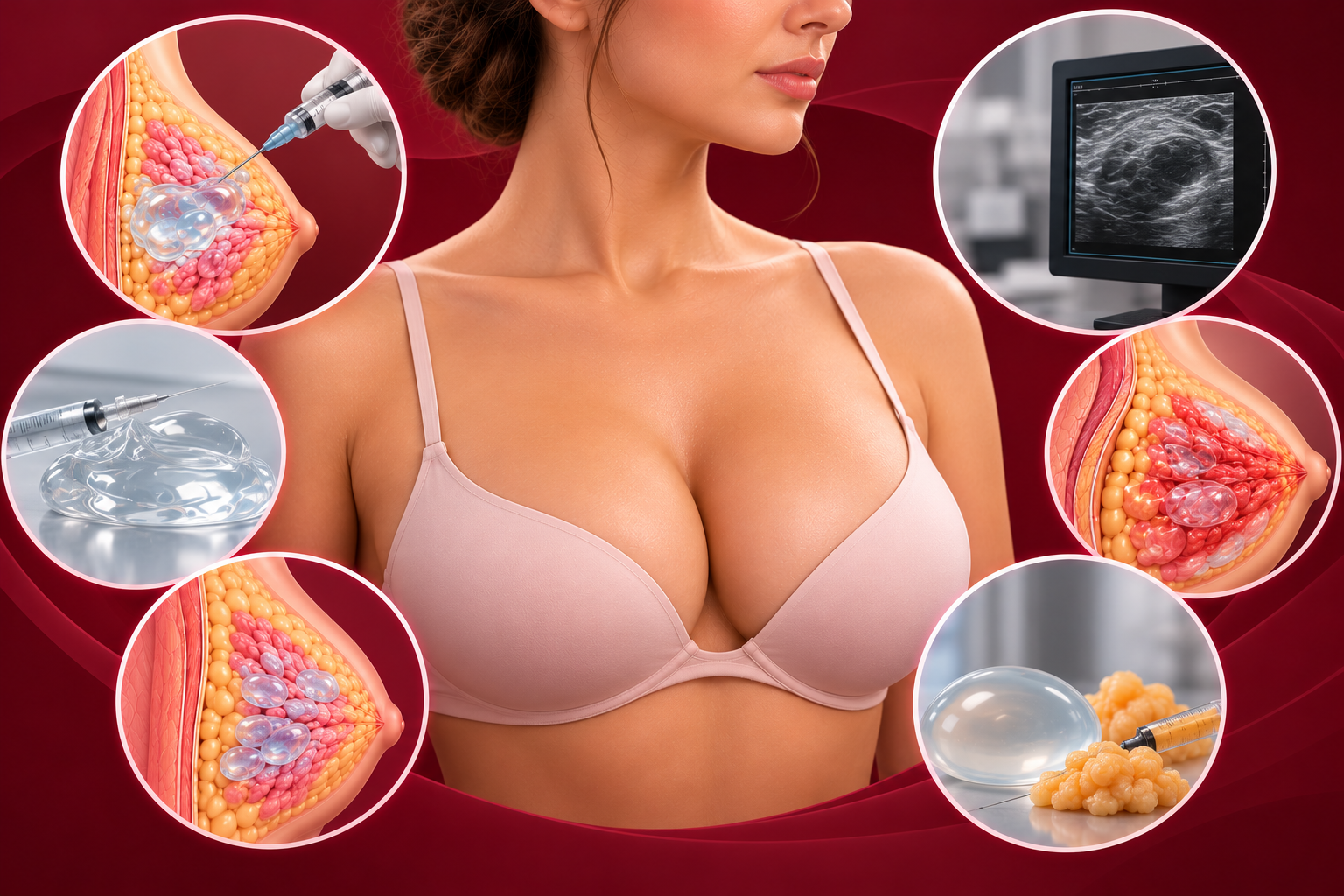
Breast screening is an important health check. Injected material can complicate it. This page is part of our guide to breast fillers.
Appearances depend on the product, how long ago it was injected and the tissue reaction.
Injected material may be mistaken for a lump or other abnormality, leading to extra tests or biopsies. It could also obscure a genuine abnormality.
If a biopsy is needed, say that you have had injections. Sampling through injected material needs careful planning.
Say so. Imaging and, if needed, tissue sampling can help characterise it.
A provider should discuss screening implications before any breast injection and record the details for future reference. See breast enlargement safety for wider pre-treatment checks.
Always tell imaging teams about breast injections. See breast enlargement safety.
Continue reading
Return to the full category guide.
Candidacy, risks and when to seek medical help.
Study findings, limitations and original publications.
The parent guide to evidence, approval and risk.
Lumps, infection and other problems to watch for.
Assessment and treatment of persistent material.